
Our laboratory is equipped with state-of-the-art facilities for cell culture, 3-D (bio)printing, and experimental research. We also provide a core facility for Micro-Computed Tomography (micro-CT) for internal (UiB) and external users.
STATE OF THE ART EQUIPMENT THAT WILL HELP
ACHIEVE OUR GOALS
Facilities
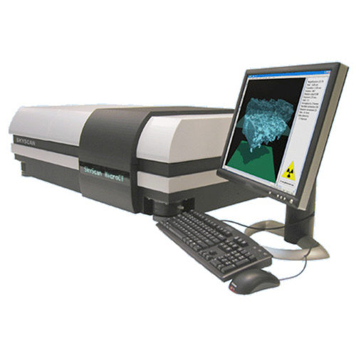
MicroCT equipment: Skyscan 1172
Computer Tomography is a non-destructive inspection technique based on differences in absorption/attenuation of X-Rays through the material. The CT-technique provides a 3D-data set of in the sample. The attenuation of X-Rays depends on the atomic number, density and thickness of the material and on the energy of the X-Ray beam. Non-destructive inspection of internal structure, tomodensity analysis, size and volumetric measurements are possible on the 2D as well as the 3D-images. The Skyscan 1172 is a high-resolution desktop X-ray micro-CT system for small samples.
Specifications for the Skyscan 1172:
- Detail detectability: <1 µm;
- Low contrast resolution (10% MTF): 5 µm;
- Pixel size at maximum magnification: 0.7 µm to 25 µm;
- Continuously variable from < 1 µm to 25 µm;
- Spot size < 5µm @ 4W;
- X-ray source: 20-100 kV, 10w (0-250 µAmax).
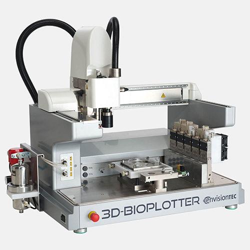
3D-Bioplotter® System
As 3D printing technology is widely applied in the field of tissue engineering, 3D-Bioplotter system is one of the most advanced product in the market. 3D-Bioplotter® System is a versatile rapid prototyping tool for processing a great variety of biomaterials for computer-aided tissue engineering (CATE), from 3D CAD models and patient CT data to the physical 3D scaffold with a designed and defined outer form and an open inner structure. The 3D-Bioplotter® has the capacity of fabricating scaffolds using the widest range of materials of any singular rapid prototyping machine, from soft hydrogels over polymer melts up to hard ceramics. Complex inner patterns can easily be designed using the 3D-Bioplotter® software to control the mechanical properties, increase cell adhesion, as well as improve the flow of nutrient media throughout the interconnecting pores of the printed implants.
The detailed information, please visit their webpage: https://envisiontec.com/3d-printers/3d-bioplotter/.
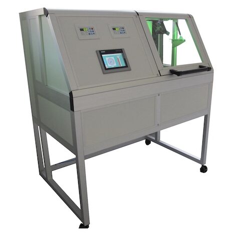
A customized perfusion bioreactor system made by Fraunhofer IGB
Bone development is a multistep process that involves the complex interplay of multiple cell types as well as local microenvironments. Bioreactor systems facilitate three-dimensional (3D) cell culture by coping with limitations of static cultivation techniques. To allow for the investigation of proper bone cell development in vitro, a mechanical stimuli plays a pivotal role in influencing cell fate during skeletal development. At physiological conditions, bone cells generally reside in a dynamic loading environment. This perfusion bioreactor system is very flexible to set a different mechanical loading, oxygen condition, shear stress with a regular condition control at either 2D or 3D culturing.
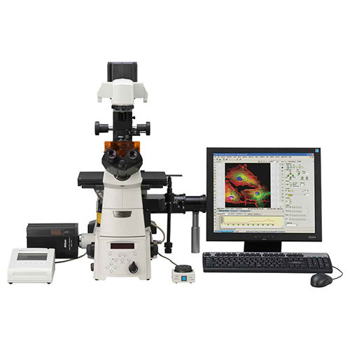
Inverted Nikon Ti Eclipse TIRF Microscope
Total Internal Reflection Fluorescence (TIRF) microscopy combines confocal-like contrast with the sensitivity of widefield microscopy for visualising of near-membrane structures. TIRF microscopy angles the illumination so that instead of passing through the sample, it reflects internally between the objective and the coverslip and then back into the objective. Only the part of the sample that is in close contact with the coverslip is illumination by an effervescent wave generated from the coverslip and extending up to ~150nm into the sample. The result is the ability to visualize membrane bound proteins, receptors, vesicles and the cortical cytoskeleton without the problems of out of focus-light as the rest of the cell is not illuminated. The combination of better sensitivity and limited illumination make this a much better choice for near-membrane imaging that confocal microscopy, especially when dealing with weakly fluorescent or easily bleached samples. The Nikon Eclipse Ti TIRF microscope combines an extremely sensitive Andor EM-CCD camera with laser-based illumination and a motorised TIRF mirror to enable true multi-colour TIRF imaging.
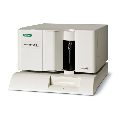
Bio-Plex® 200 Systems
The Bioplex 200 system can analyze immunoassays, complex genetic analysis, and enzymatic assays in one format. The reactants, (antibodies, oligonucleotides, substrates, etc.) are attached to unique fluorescent microscopic beads (microspheres). The unique fluorescent emission for a given microsphere enables the Bioplex to identify discrete measurements of multiple microsphere based reactions from a single sample. xMAP technology allows multiplexing of up to 100 unique assays within a single sample, both rapidly and precisely
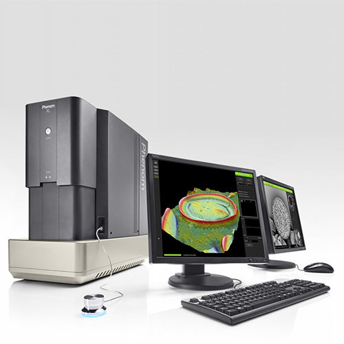
Phenom XL Scanning Electron Microscope
The Phenom XL Scanning Electron Microscope (SEM) pushes the boundaries of compact desktop SEM performance. It features the proven ease-of-use and fast time-to-image of any Phenom system.
It is equipped with a chamber that allows analysis of large samples up to 100 mm x 100 mm. A proprietary venting/loading mechanism ensures the fastest vent/load cycle in the world, providing the highest throughput.
A newly developed compact motorized stage enables the user to scan the full sample area, and yet the Phenom XL is a desktop SEM that needs little space and no extra facilities.
Equipment Booking
For internal users at UiB, the price is 400 kr per hour for scanning and 500 kr per hour with assistance for analysis.
For external users, the price is 600 kr per hour for scanning and 700 kr per hour with assistance for analysis.
Biological lab leader: Prof. Kamal Mustafa booking@tissueengineering.no